


| Story Views | |
| Now: | |
| Last Hour: | |
| Last 24 Hours: | |
| Total: | |
Ultrasound Scalpel Gently Destroys Liver Tumors

Focused ultrasound can effectively destroy tumor cells. Until now, this method has only been used for organs such as the prostate and uterus. At the European Congress of Radiology, Fraunhofer researchers will present a method, developed as part of the TRANS-FUSIMO EU project, that enables focused ultrasound treatment of the liver, an organ that moves while breathing. In the future, this could enable treatment of certain liver tumors in a more gentle way.
Ultrasound has long served as a diagnostic method. Its application as a form of therapy treatment, however, is relatively new. In this process, ultrasound waves are highly concentrated to destroy diseased tissue, tumor cells in particular, and render them harmless. Focused ultrasound benefits patients in several ways. The therapy is completely non-invasive and can be performed without anesthesia, and there are no operation wounds.
Doctors wish to use focused ultrasound to treat tumors in moving organs, such as the liver, shown here.
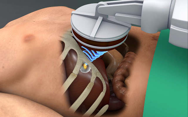
Photo Fraunhofer MEVIS
Until now, however, the method has only been approved for a limited number of indications, such as treatment of prostate cancer, bone metastases, and uterine myoma. To treat organs that move when patients breathe, the method can only be partially applied. Doctors have to rely on patients to hold their breath or put them under anesthesia, so they can control the patient’s breath.
The scientists working in the TRANS-FUSIMO EU project, coordinated by the Fraunhofer Institute for Medical Image Computing MEVIS in Bremen, are following another path. They refocus the ultrasound beam to the movement of the liver to reach the tumor effectively while sparing the surrounding healthy tissue. The fundamental technology needed for the method is now ready. The researchers presented important preliminary results as part of an industry symposium at the European Radiology Congress (ECR) in Vienna on March 1.
In this therapy concept, the patient lies in an MRI scanner during the procedure. Every tenth of a second, the scanner produces an image showing the current position of the liver. The ultrasound transducer, a device equipped with more than 1000 small ultrasound transmitters, sits on the patient’s stomach. These can be directed so that their waves converge precisely at a point as small as a grain of rice. There, they unleash their destructive effect – the tumor cells become completely cooked. The MRI scanner controls the process, measuring the temperature in the liver and ensuring that the correct spots are sufficiently heated.
Real-time software that can glance into the immediate future
Project manager Sabrina Haase, mathematician at Fraunhofer MEVIS, explains the problem. “Generating an image of the liver’s position every tenth of a second is not fast enough to reliably direct the ultrasound beam. This is why we developed software that can see into the immediate future and calculate the next position of the treated region.” The program determines the path for the focused ultrasound waves to reach the liver tumor even when the patient moves while breathing. Developing the software was quite challenging: it must run both highly reliably and in real time.
Another difficulty facing the scientists was the fact that the ribs lie in front of the liver. To prevent the beams from damaging the ribs, elements in the ultrasound transducer that would have hit the ribs were deactivated, much like blocking the holes in a showerhead which spray water in an unwanted direction.
“We have completed the technical development phase and have already run preliminary tests,” says Haase. In the test, a robotic arm moved a gel model back and forth in the MR scanner to simulate the liver movement inside the body. At the same time, the gel phantom was exposed to focused ultrasound, and the MRI scanner monitored the temperature distribution. “The results match our expectations,” says Haase. “Now, we can pursue the next steps.”
The first TRANS-FUSIMO tests on patients are planned for mid-2018. Thereafter, in cooperation with an industry partner, medical product certification can be tackled. If the method proves itself, in the future, it would be possible to treat other organs that move with breathing, such as the kidney, pancreas, or even lungs.
The TRANS-FUSIMO (“Clinical Translation of Patient-Specific Planning and Conducting of FUS Treatment in Moving Organs”) EU project began in the beginning of 2014 and runs until the end of 2018. Eleven institutions form seven different countries participate in the project. In addition to clinics and universities, participants include medical technology companies. The Fraunhofer Institute for Medical Image computing in Bremen coordinates the project, which is funded with 5.6 million Euros. For further information,
Contacts and sources:
Bianka Hofmann
Fraunhofer Institute for Medical Image Computing MEVIS
