


X Rays and Anoles

An exciting week in the Revell Lab, we received our order of 20 poles from Cabelas, and I picked up our new custom portable x-ray system in Newark yesterday.
The use of x-ray technology has been mention previously in AA- here, here , here, here, and here. The Losos Lab has used a similar portable x-ray system for the last several years with great success, and so we have obtained our own unit. One of the great advantages of these systems is that they allow researchers to gather highly detailed morphological data without harming the lizards and without using tedious methods such as dissection. The animals are simply anesthetized, imaged, and released after recovery. The Revell Lab has grand aspirations for our system- our graduate student Kristin Winchell plans to use it this summer in her studies of Anolis urban ecology.
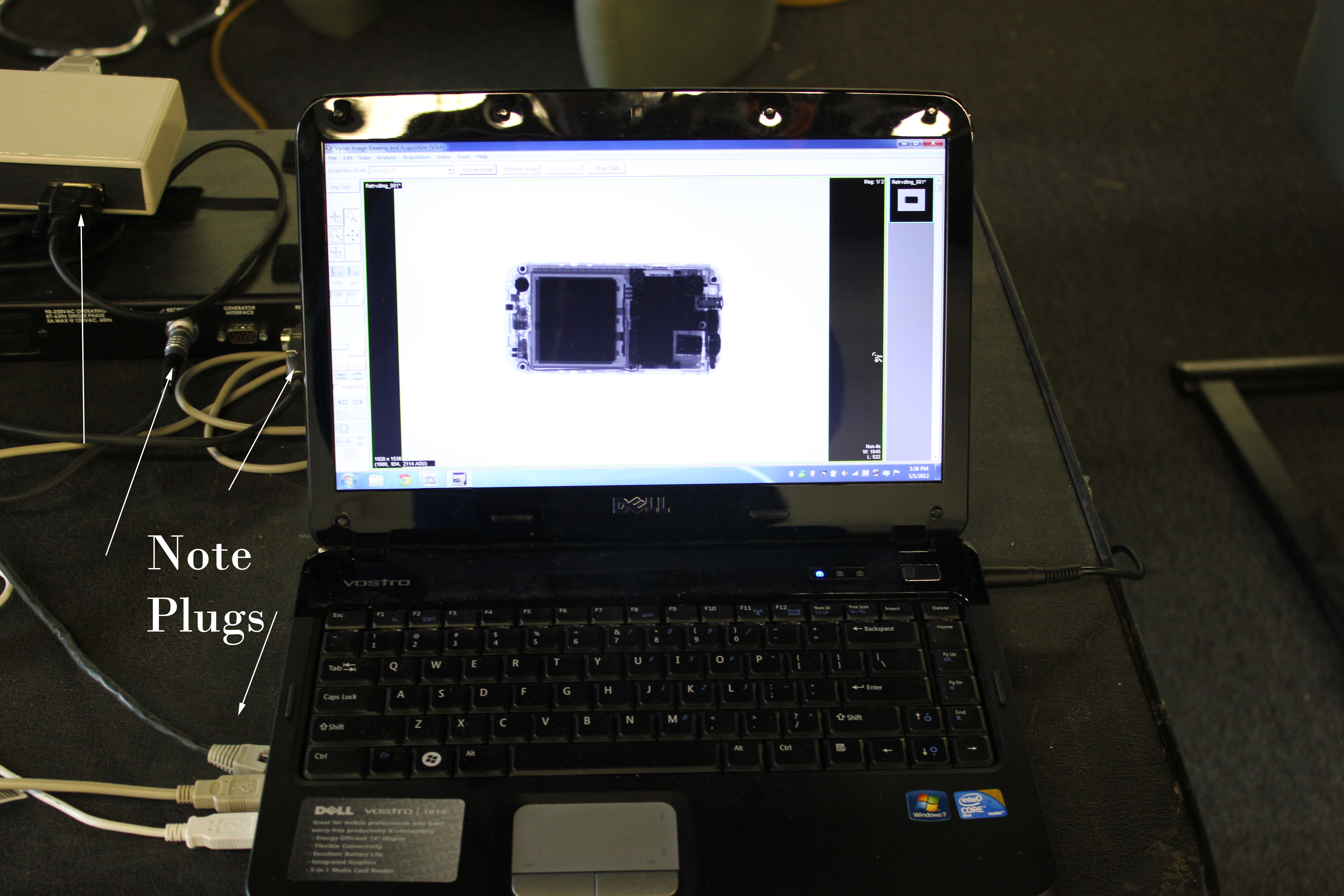
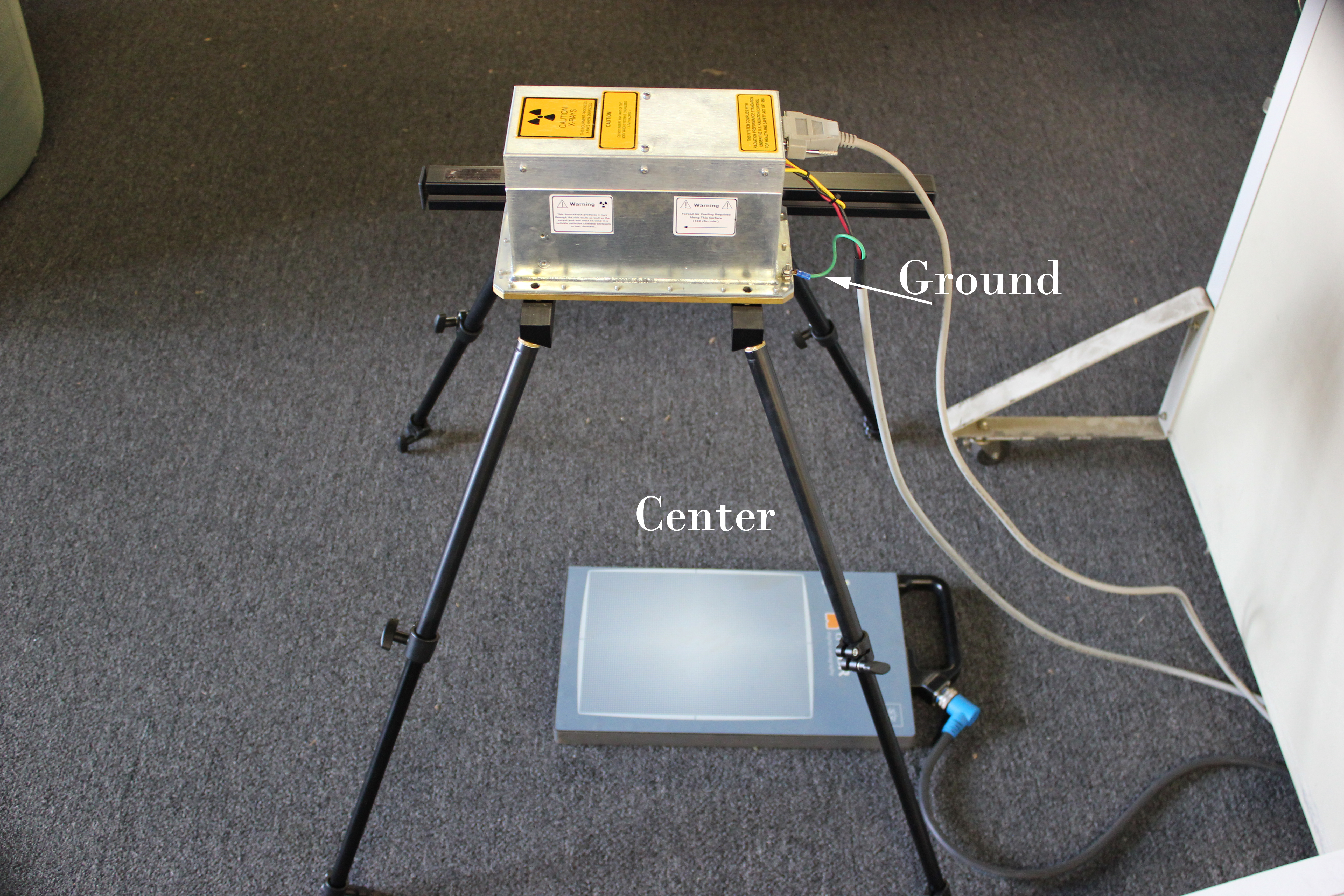
The system is fairly simple and easy to use. Below is a picture of the setup, which includes an x-ray source (above) and a digital detector plate (below). A 40 degree beam is produced when the source is engaged from a remote control system consisting of a control box, power supply, and control software on a laptop. At 9.6 x 7.7 inches, our PaxScan detector plate is large enough to image even the crown giants.
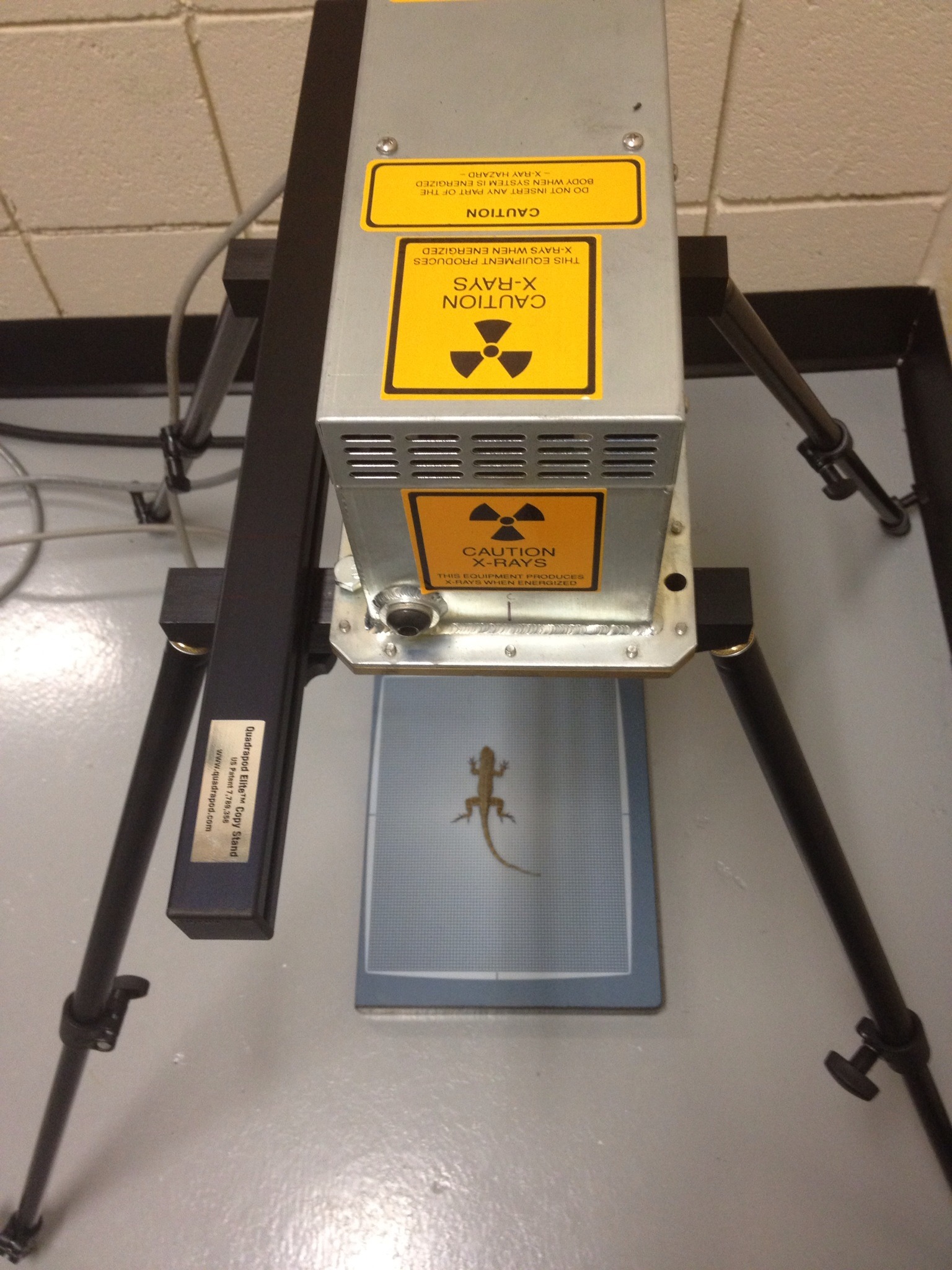
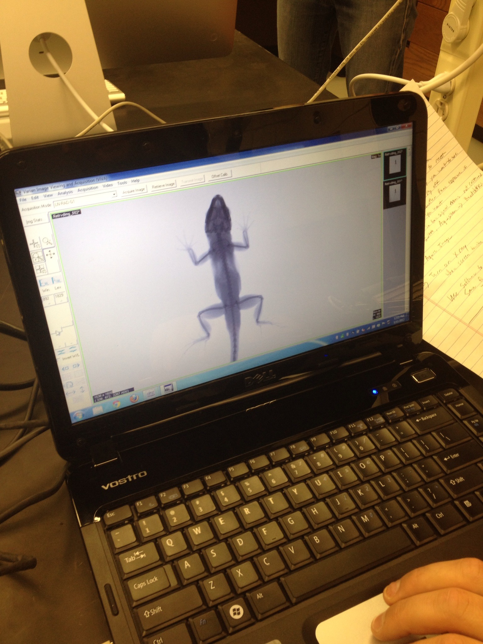
Below is an example of an (anesthetized) adult male A. cristatellus, an animal that was born in captivity at Harvard and now resides comfortably at UMass Boston. In fact, as Liam observed, today’s photo session was probably the most excitement he has had in quite a while!

Finally, the best feature of this system is that it all packs neatly into a Pelican case:

Source:
