


| Story Views | |
| Now: | |
| Last Hour: | |
| Last 24 Hours: | |
| Total: | |
Needle in a haystack search turns up compound that might mend broken hearts

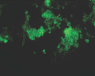 |
| Stem cell-derived heart muscle cells |
The first task of developing a stem cell-based therapy is coaxing the stem cells to turn into the desired cell type – neurons for brain diseases, pancreatic cells for diabetes, or, in the case of some recent work by a group of CIRM grantees, heart muscle cells for heart disease.
A group of researchers from Sanford-Burnham and Human BioMolecular Research Institute in San Diego screened 17,000 compounds to find one that would push embryonic stem cells from mice to form heart tissue. They published the results of that screen–which produced one compound that could push stem cells to form heart tissue–today in the journal Cell Stem Cell.
A press release from Sanford-Burnham quotes researcher Mark Mercola, who is director of Sanford-Burnham’s Muscle Development and Regeneration Program and senior author of the study:
“Heart disease is the leading cause of death in this country. Because we can’t replace lost cardiac muscle, the condition irreversibly leads to a decline in heart function and ultimately death. The only way to effectively replace lost heart muscle cells—called cardiomyocytes—is to transplant the entire heart. Using a drug to create new heart muscle from stem cells would be far more appealing than heart transplantation.”
The way scientists go about screening all these compounds is essentially a high-tech method of extracting a needle from a molecular haystack. They put embryonic stem cells within small indents on lab plates, each of which had 384 such indents called wells. Then, using a robot arm to automate the process, they put a different compound in each of the wells. The stem cells then bathed in those different compounds for a few days. Some of the compounds killed the cells outright, while others nudged the cells to begin forming different kinds of tissues. Since the goal of this particular screen was to find those cells that had begun to form heart tissue, the scientists had engineered the cells to turn green when a gene involved in heart formation was active. When the group ran those plates through a special sensor a few days later, all they had to do was look for wells containing green cells. They found one such group of cells, corresponding to one of the compounds.
The group is now working with San Diego biotech company ChemRegen to turn the compound into a drug that could be used to repair damaged heart tissue.
Here’s a video Sanford-Burnham produced about the work:
CIRM Funding: Erik Willems (T2-00004), John Cashman (RS1-00169-1), Mark Mercola (RC1-00132)
A.A.
![]() Erik Willems, Joaquim Cabral-Teixeira, Dennis Schade, Wenqing Cai, Patrick Reeves, Paul J. Bushway, Marion Lanier, Christopher Walsh, Tomas Kirchhausen, Juan Carlos Izpisua Belmonte, John Cashman, & Mark Mercola (2012). Small Molecule-Mediated TGF-β Type II Receptor Degradation Promotes Cardiomyogenesis in Embryonic Stem Cells Cell Stem Cell DOI: 10.1016/j.stem.2012.04.025
Erik Willems, Joaquim Cabral-Teixeira, Dennis Schade, Wenqing Cai, Patrick Reeves, Paul J. Bushway, Marion Lanier, Christopher Walsh, Tomas Kirchhausen, Juan Carlos Izpisua Belmonte, John Cashman, & Mark Mercola (2012). Small Molecule-Mediated TGF-β Type II Receptor Degradation Promotes Cardiomyogenesis in Embryonic Stem Cells Cell Stem Cell DOI: 10.1016/j.stem.2012.04.025
Read more stem cell research news from the California Institute for Regenerative Medicine by visiting our blog at cirmresearch.blogspot.com.
Learn more about the California Institute for Regenerative Medicine
2012-08-03 06:45:33
Source: http://cirmresearch.blogspot.com/2012/08/needle-in-haystack-search-turns-up.html
Source:
